Introduction:
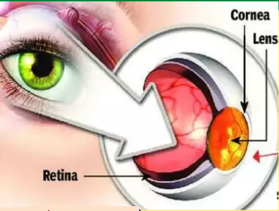
Retina is the light sensitive layer of tissue that lines the inside of the eye and sends visual messages through the optic nerve to the brain. When the retina detaches, it is lifted or pulled from its normal position. If not treated promptly, retinal detachment can cause permanent vision loss.
(PLEASE CLICK FOR TAMIL IMAGE FORMAT:RETINAL DETACHMENT )
Symptoms:


• Floaters: black dots or stars that move through your field of vision.
• Sudden, short flashes of light.
• Blurry vision.
• Curtain-shaped vision loss.
Causes :


• Extremely nearsighted.
• Had a retinal detachment in the other eye.
• Family history of retinal detachment.
• Had an eye injury.
• Has had cataract surgery.
Risk factors:


• Severe myopia.
• Eye injury or cataract surgery.
• Family history of retinal detachment.
• Lattice degeneration (Weakness in the edges of your retina).
• Diabetic retinopathy (damage to the blood vessels in your retina due to diabetes)
• Age.
• Posterior vitreous detachment (Vitreous gel in your eye moves away from your retina.
Types of retinal detachment:
Rhegmatogenous :

• This is the most common type.
• It occurs due to Retinal holes.
• Retinal degeneration, or Injury to retina.
• Surgery
• Due to age vitreous gel moves away from your retina. This can produce Retinal Detachment . if your retina is weak.
Traction:
This type occurs when new tissue forms and pulls the normal Retina.
Exudative:
This occurs when the retinal fluid forms in your retina. The fluid pushes your retina away from the tissues behind it. This can happen due to injury age related nerve damage or Injection.
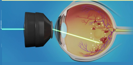
Laser cryopexy:
Both methods of laser thermal or freezing cryopexy can be done if detected early.
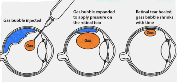
Pneumatic Retinopexy
It works well for small and easily closable retinal detachment.
Your doctor injects a small gas bubble into your Vitreous gel. It presses on the top of your retina and pushes the nerve into its proper place.
You need to keep your head in a certain position for several days to keep the bubble in the right place.
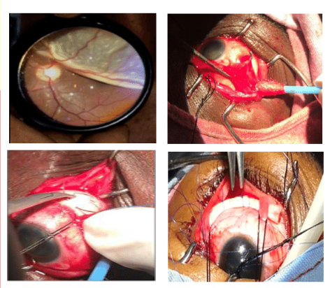
(Scleral Buckle)
Scleral buckle is the solution to retinal detachment. In a surgery called Scleral Buckling, a small synthetic band is placed outside the Sclera. This is used to push the retina slightly inwards. So that it goes in and touches and reattaches to the detached part.
Vitrectomy:
Although up to 90% of patients with retinal detachment are successfully treated with modern therapy, although sometimes re-treatment may be required, it is not possible to predict improvement in vision. Vision improvement is more likely to occur if treatment is done easily.
.png)


.jpg)


Comments